太阳成集团tyc151com(中国)搜狗百科

NEWS

  • 02 11
       The research team led by Prof. Hao Wang has recently published an article titled “Neural adaption in midbrain GABAergic cells contributes to high-fat-diet induced obesity” in Science Advances.In modern society, high-calorie food is readily available and easily accessible, leading to a steady increase in the incidence of obesity. Obesity, in turn, contributes to the rise of other related diseases such as hypertension, hyperlipidemia, and diabetes, placing a significant burden to both society and families. Consequently, obesity has emerged as a pressing public health concern worldwide, with limited treatment options.While previous studies have demonstrated that weight control can be achieved through modifications in dietary structure and lifestyle habits, it is often observed that individuals in this population tend to regain weight within five years. This phenomenon may be attributed to the impact of high-calorie foods, which not only influence body weight and metabolism, but also induce irreversible changes in the central nervous system. To delve deeper into this issue, Professor Hao Wang and his team from Zhejiang University’s School of Brain Science and Brain Medicine conducted a research study entitled “Neural adaption in midbrain GABAergic cells contributes to high-fat-diet induced obesity”, which was published in Science Advances. The study discussed the neural adaptations observed in midbrain GABAergic cells as a result of high-fat-diet (HFD) induced obesity.Professor Hao Wang's research team has been dedicated to investigating the neural circuit mechanisms involved in regulating energy metabolic homeostasis. In their previous work published in Cell Reports (2019), they made a significant discovery that GABAergic neurons in the ventrolateral periaqueductal grey (vlPAG) region possess an appetite-suppressing effect.In their current study, the team utilized a chemogenetic approach to activate vlPAG GABAergic neurons and observed a reversal of the obesity phenotype in high-fat-diet-induced obese (DIO) mice. This rescue effect was achieved by reducing 24-hour food intake, increasing energy metabolism levels, and inducing browning of adipose tissue.Through the use of in vivo fiber-photometry calcium imaging, the researchers discovered that calcium signals originating from vlPAG GABAergic neurons are suppressed during food intake. Notably, these neurons exhibited stronger suppression in DIO mice. Further electrophysiological recordings provided insights into the mechanisms underlying these observations. The reduced excitability of the "food-suppressed" neurons in obese mice was found to be a result of increased presynaptic inhibitory inputs and a decrease in intrinsic excitability of the neurons themselves. Consequently, chronic high-fat food intake leads to long-term inhibition of these "food-suppressor" neurons, ultimately contributing to increased food intake and obesity.The team further employed single-cell nuclear transcriptome sequencing technology to conduct a comprehensive analysis of gene expression changes in GABAergic neurons within the vlPAG of obese mice, comparing them with control mice. They identified a crucial gene called CACNA2D1, which exhibited significantly reduced expression levels in obese mice. To investigate the potential role of CACNA2D1, the team performed AAV-overexpression of CACNA2D1 in the vlPAG of obese mice. Remarkably, this intervention led to the rescue of the obesity phenotype observed in DIO mice. The rescue effect was achieved by reducing food intake and promoting adipose tissue browning. Additionally, the restoration of CACNA2D1 expression resulted in the recovery of excitability in the "food-suppressor" neurons located in the vlPAG. These findings suggest that CACNA2D1 holds promise as a potential target for the treatment of stubborn obesity.  In summary, Prof. Hao Wang's team found that the "food-suppressor" neurons in the vlPAG are involved in the regulation of energy balance and help maintain body weight homeostasis. However, long-term high-fat food intake will cause these "food-suppressor" neurons to go on strike, which makes the animals unable to stop eating high-fat food, and the vicious cycle of excessive food intake will continue. CACNA2D1 may be a potential target for the treatment of recalcitrant obesity.GABAergic neurons in the periaqueductal grey regulate weight metabolic homeostasis. Professor Hao Wang from the School of Brain Science and Brain Medicine at Zhejiang University served as the corresponding author, while Dr. Xiaomeng Wang and Dr. Xiaotong Wu were the co-first authors of this paper. The study received support from Professor Shumin Duan, Professor Xiaoming Li, Professor Yudong Zhou, Professor Chen Li, Professor Han Xu, Professor Jiadong Chen, Professor Wei Gong, Professor Fang Guo, and Professor Ruimao Zheng. Additionally, Dr. Bingwei Wang, along with PhD students Hao Wu and Hanyang Xiao, made significant contributions to this research. Funding for this study was provided by the National Natural Science Foundation of China and Boehringer Ingelheim in Germany.
    view details
  • 19 10
    The research team led by Prof. Hailan Hu has recently published an article titled Sustained Antidepressant Effect of Ketamine through NMDAR Trapping in the LHb on Nature online on Oct 18th, 2023. This research revealed ketamine trapped in NMDAR to mediate the mechanism of the sustained antidepressant effects of ketamine.The use-dependent trapping properties of ketamine for NMDAR are the essence of its sustained antidepressant effects. Ketamine, an N-methyl-d-aspartate receptor (NMDAR) antagonist, has revolutionized the treatment of depression because of its potent, rapid and sustained antidepressant effects. Although the elimination half-life of ketamine is only 13 min in mice, its antidepressant activities can last for at least 24 h. This large discrepancy poses an interesting basic biological question and has strong clinical implications. Here we demonstrate that after a single systemic injection, ketamine continues to suppress burst firing and block NMDARs in the lateral habenula (LHb) for up to 24 h. This long inhibition of NMDARs is not due to endocytosis but depends on the use-dependent trapping of ketamine in NMDARs. The rate of untrapping is regulated by neural activity. Harnessing the dynamic equilibrium of ketamine–NMDAR interactions by activating the LHb and opening local NMDARs at different plasma ketamine concentrations, we were able to either shorten or prolong the antidepressant effects of ketamine in vivo. These results provide new insights into the causal mechanisms of the sustained antidepressant effects of ketamine. The ability to modulate the duration of ketamine action based on the biophysical properties of ketamine–NMDAR interactions opens up new opportunities for the therapeutic use of ketamine.  Activating LHb bidirectionally modulates the duration of the ketamine antidepressant effect  Website: https://doi.org/10.1038/s41586-023-06624-1 HAILAN HU'S RESEARCH GROUP: For social animals, emotions and health are regulated by various social behaviors. Hailan Hu's group is dedicated to studying the neural basis and plasticity mechanisms of emotion and social behavior. They use cutting-edge techniques including imaging, electrophysiology (both in vitro and in vivo), molecular genetics, and optogenetics to conduct deep analysis of emotion- and social behaviors- and their related neural circuits.  
    view details
  • 03 10
    The research team led by Prof. Hailan Hu has recently published an article titled Stress Relief as a Natural Resilience Mechanism against Depression-like Behaviors on Neuron online on Sep 29th, 2023. This research discovered relief as a natural resilience mechanism against depression, and deconstructed the neural circuit mechanisms underlying relief.Unraveling relief as a homeostatic defense mechanism for mood regulation and clarifying the underlying neural circuit mechanismRelief, the appetitive state after the termination of aversive stimuli, is evolutionarily conserved. The inherent opponency in the valence between stress and relief raises the intriguing possibility that relief may counteract the detrimental effects of stress, playing a role in stress resilience. However, such a possibility has not been tested experimentally. Understanding the behavioral role of this well-conserved phenomenon and its underlying neurobiological mechanisms are open and important questions.Based on the correlative discovery that relief magnitude strongly correlates with resilience level to depression, researchers further revealed that blocking stress relief causes vulnerability to depression-like behaviors, whereas natural rewards supplied shortly after stress promotes resilience. Stress relief is mediated by reward-related mesolimbic dopamine neurons, which show minute-long, persistent activation after stress termination. Circuitry-wise, activation or inhibition of circuits downstream of the ventral tegmental area during the transient relief period bi-directionally regulates depression resilience. These results reveal an evolutionary function of stress relief in depression resilience, and identify the neural substrate mediating this effect. Importantly, our data suggest a behavioral strategy of augmenting positive valence of stress relief with natural rewards to prevent depression.Website: https://www.cell.com/neuron/fulltext/S0896-6273(23)00668-2Figure. Physiological utility of relief and underlying dopaminergic circuit mechanisms
    view details
  • 12 09
    The research team led by Prof. Xiao-Ming Li has recently published an article titled Distinct Circuits from Central Lateral Amygdala to Ventral Part of Bed Nucleus of Stria Terminalis Regulate Different Fear Memory in Biological Psychiatry on Sep 5th, Beijing time.This research established a functional role for distinct central lateral amygdala to ventral part of stria terminalis circuits in the differential regulation and appropriate maintenance of fear.The ability to differentiate stimuli predicting fear is critical for survival, however, the underlying molecular and circuit mechanisms remain poorly understood. This work identified the projections from central lateral amygdala (CeL) protein kinase C δ (PKCδ) positive neurons and somatostatin (SST) positive neurons to the ventral part of bed nucleus of stria terminalis (vBNST) GABAergic and glutamatergic neurons. Prolonged optogenetic activation or inhibition of PKCδCeL-vBNST pathway specifically reduced context fear memory, whereas SSTCeL-vBNST pathway mainly reduced tone fear memory. Intriguingly, optogenetic manipulation of vBNST neurons received the projection from PKCδCeL exerted bidirectional regulation of context fear, whereas manipulation of vBNST neurons received the projection from SSTCeL neurons could bidirectionally regulate both context and tone fear memory. The presence of δ and κ opioid receptor protein expression within the CeL-vBNST circuits potentially accounted for the discrepancy between prolonged activation of GABAergic circuits and inhibition of downstream vBNST neurons. Finally, administration of an opioid receptor antagonist cocktail on the PKCδCeL-vBNST or SSTCeL-vBNST pathway successfully restored context or tone fear memory reduction induced by prolonged activation of the circuits. This study provides the first evidence that distinct extended amygdala circuits participated in differently regulating fear memory, which can pave the way for an innovative approach to drug development for a range of fear-related syndromes.Pro. Xiao-Ming Li from Zhejiang University School of Medicine is the main corresponding author. Dr Yi Zhu, PhD candidates Shi-Ze Xie and Ai-Bing Peng are the first authors. This study was funded by the National Natural Science Foundation of China, STI2030-Major Projects, Key-Area Research and Development Program of Guangdong Province, Key R&D Program of Zhejiang Province, Fundamental Research Funds for the Central Universities, CAMS Innovation Fund for Medical Sciences, Innovative and Entrepreneur Team of Zhejiang for 2020 Biomarker-Driven Basic and Translational Research on Major Brain Diseases and the fellowship of China Postdoctoral Science Foundation.
    view details
  • 27 07
    The research team led by Prof. Hongbin Yang has recently published an article titled Dopamine Release and Negative Valence Gated by Inhibitory Neurons in the Laterodorsal Tegmental Nucleus in Neuron on July 26, 2023, Beijing time. This research uncovered separate GABAergic subpopulations in a single brainstem nucleus that relay unpleasant stimuli to the mesolimbic DA system through direct and indirect projections, which is critical for establishing a circuit-level understanding of how negative valence is encoded in the mammalian brain. GABAergic neurons in the laterodorsal tegmental nucleus (LDTGABA) encode aversion by directly inhibiting mesolimbic dopamine (DA). Yet, the detailed cellular and circuit mechanisms by which these cells relay unpleasant stimuli to DA neurons and regulate behavioral output remain largely unclear. Here, this research shows that LDTGABA neurons bidirectionally respond to rewarding and aversive stimuli in mice. Activation of LDTGABA neurons promotes aversion and reduces DA release in the lateral nucleus accumbens. Furthermore, this research identified two molecularly distinct LDTGABA cell populations. Somatostatin-expressing (Sst+) LDTGABA neurons indirectly regulate the mesolimbic DA system by disinhibiting excitatory hypothalamic neurons. In contrast, Reelin-expressing LDTGABA neurons directly inhibit downstream DA neurons.  Prof. Hongbin Yang from Zhejiang University MOE Frontier Science Center for Brain Science & Brain-Machine Integration is the corresponding author. Yonglan Du, Siyao Zhou, Chenyan Ma and Hui Chen are co-first authors. Ana Du, Guochuang Deng, Yige Liu also made significant contributions. This research was strongly supported by Prof. Shumin Duan and Prof. Lammel. This work was supported by grants from the STI2030-Major Projects, National Natural Science Foundation of China, NSFC-Guangdong Joint Fund-U20A6005, Key R&D Program of Zhejiang province, Fund for Medical Science and Key R&D Program of Guangdong Province, and the Fundamental Research Funds for the Central Universities. 原文链接:https://www.cell.com/neuron/fulltext/S0896-6273(23)00480-4
    view details
  • 11 07
    AbstractOn July 7th, 2023, Xuhua Wang's research group at Zhejiang University School of Medicine published a study titled Porous microneedle patch with sustained extracellular vesicles delivery mitigates severe spinal cord injury in Nature Communications. This team fabricated a porous microneedle patch based on mesenchymal stem cells (MN-MSC) containing mesenchymal stem cells and microneedle arrays for delivering extracellular vesicles to spinal cord injury sites for treatment. The microneedle array with mechanical strength matching that of the spinal cord can prevent damage to the surrounding spinal cord tissues. The porous microstructure of the microneedles facilitates extracellular vesicle delivery. Mesenchymal stem cells can continuously provide extracellular vesicles. This study provides new insights into the treatment of spinal cord injury.The extracellular vesicles secreted by mesenchymal stem cells have become a promising treatment to inhibit neural inflammation triggered by spinal cord injury. However, how to effectively deliver extracellular vesicles to the spinal cord injury site while minimizing damage to the surrounding tissues or axons remains a challenge. Therefore, a patch was proposed for treating spinal cord injury through sustained delivery of extracellular vesicles. This team evaluated the efficacy of a porous microneedle patch based on mesenchymal stem cells in a rat model of spinal cord injury and found it could improve the microenvironment in the injured tissue, promote angiogenesis, and protect residual neural tissues. Importantly, sustained delivery of extracellular vesicles for at least 7 days led to significant functional recovery. The porous microneedle patch based on mesenchymal stem cells developed by this team brings hope for the rehabilitation of patients with spinal cord injury.Research backgroundSpinal cord injury is a central nervous system trauma caused by trauma, with an estimated global incidence of 15-40 case per million people. This results in an annual global economic burden of up to 40 billion dollars [1]. Repairing spinal cord injuries remains a key challenge, as currently no method can completely cure them. Various approaches exist to repair and improve spinal cord injury outcomes, including the use of stem cells, particularly mesenchymal stem cells (MSCs). Studies demonstrate MSCs have a therapeutic effect on spinal cord injuries, especially via their secreted extracellular vesicles (EVs), which are thought to underlie the therapeutic benefits. A disadvantage of intrathecal administration of MSCs or EVs is that most migrate into the blood or other tissues, with very few reaching the central nervous system. Recently, immobilizing EVs in peptide-modified hydrogels was shown to improve spinal cord injury treatment versus traditional delivery methods by increasing EV efficiency [2].However, the delivery efficiency and duration of this hydrogel method are insufficient to exert the optimal therapeutic effect [3]. Treating the neural inflammation and apoptosis caused by spinal cord injury requires sustained delivery for at least 1 week. Unfortunately, this hydrogel method cannot provide sustained delivery of adequate extracellular vesicles over this timeframe. Addressing this limitation is an urgent problem needing solved in spinal cord injury treatment. Furthermore, directly transplanting mesenchymal stem cells or extracellular vesicles into the spinal cord injury site risks damaging surrounding healthy tissues, potentially causing unpredictable side effects [4]. Therefore, developing a device that can efficiently and continuously supply extracellular vesicles over an extended period plays a crucial role in treating acute spinal cord injury.Research resultsAs shown in Figures 1 and 2, the porous microneedle patch based on mesenchymal stem cells developed in this study has good nanoscale sizes (~100 nm), biocompatibility, mechanical properties, and the ability to efficiently and sustainably deliver extracellular vesicles in vivo. In vivo, the mesenchymal stem cells can survive for a sufficiently long time at the injury site to achieve the optimal treatment time for spinal cord injury. The research team proposed this approach to improve the therapeutic effect without the need for direct intrusion of mesenchymal stem cells into the spinal cord. The microneedle patch can sustainably deliver extracellular vesicles in vivo. On day 7 after implantation, the cells still have good viability and the released extracellular vesicles can be successfully delivered to the injured area of the spinal cord, achieving sustained treatment of spinal cord injury, as shown in Figure 3.Figure 1. Schematic of implantation of the porous microneedle patch based on mesenchymal stem cells at the spinal cord injury site.Figure 2. Characterization of the porous microneedle patch based on mesenchymal stem cells.Figure 3. Sustained extracellular vesicle delivery therapy by the porous microneedle patch based on mesenchymal stem cells at 7 days after implantation.After validating the repair function of the porous microneedle patch based on mesenchymal stem cells, the team conducted a more systematic evaluation of the efficacy of the designed porous microneedle patch based on mesenchymal stem cells. In the treatment group, by comparing gene expression and tissue morphology (Figures 4-5), it was found that although MN-EV and Gel-MSC had some therapeutic effects, the rats in the MN-MSC group had more significant therapeutic effects and achieved more significant functional recovery (Figures 6-7), closer to normal rats. These results indicate that the porous microneedle patch based on mesenchymal stem cells designed in this study can exert neuroprotective effects and help rats with spinal cord injury achieve eventual motor functional recovery.This study not only developed an efficient, sustained extracellular vesicle delivery microneedle patch, but also promoted functional recovery after spinal cord injury. This provides new insights and methods for the treatment of spinal cord injury using extracellular vesicles. The results demonstrate that one of the effective methods for spinal cord injury treatment is sustained and effective delivery of extracellular vesicles to the injury site to alleviate inflammation and protect neural tissues. This study constructed a porous microneedle patch based on mesenchymal stem cells, which can continuously deliver vesicles from mesenchymal stem cells to reach the injured spinal cord tissue, avoiding the one-time loss of extracellular vesicles. The microneedle portion of the patch is made of porous GelMA hydrogel with good biocompatibility, which can remain on top of the lesion tissue for a long time and maintain sustained release until the optimal treatment time, achieving the maximum efficacy of mesenchymal stem cell vesicles.Figure 4. Gene expression analysis of treatment for spinal cord injury using the porous microneedle patch based on mesenchymal stem cells.Figure 5. Protective effects of the porous microneedle patch based on mesenchymal stem cells on spinal cord injury.Figure 6. The porous microneedle patch based on mesenchymal stem cells improves recovery of hindlimb function in rats with spinal cord injury.Figure 7. Electrophysiological experiments show that the porous microneedle patch based on mesenchymal stem cells has good control over rat muscles.Research outlookWhile the current research team has achieved promising therapeutic effects, there remains ample opportunity to further advance microneedle patch development for spinal cord injury treatment. In subsequent research, Professor Wang's team has independently developed a high optical absorption cross-linking initiator that enables more precise two-photon printing of personalized porous microneedles, with printing accuracy far surpassing that of conventional commercially available initiators. This breakthrough is anticipated to further enhance the therapeutic efficacy of microneedle patches and achieve more comprehensive functional repair.Moving forward, the team aims to continue optimizing the design and manufacturing of personalized MSC porous microneedle patches, working to successfully translate a safe and efficient spinal cord injury treatment microneedle patch to the clinic within the next few years for the benefit of more patients with spinal cord injuries.Link to original paper:https://www.nature.com/articles/s41467-023-39745-2Brief introduction of corresponding authorXuhua Wang is a researcher at the School of Brain Science and Brain Medicine at Zhejiang University. His lab primarily focuses on developing therapeutic agents and drug delivery systems for central nervous system diseases. His research interests include large language models, AI-empowered drug design, and AAV gene delivery vector engineering. More information can be found on his lab homepage at www.zjuwanglab.com. Professor Xuhua Wang from the School of Brain Science and Brain Medicine at Zhejiang University is the corresponding author. Dr. Ao Fang is the first author and the co-first authors are Yifan Wang, Guonai Yu, and Yanmig Zuo. This research was strongly supported by Prof. Xiaosong Gu from Nantong University and his team. We sincerely thank Prof. Shumin Duan from Zhejiang University for providing strong support to facilitate this project. This work was supported by the Major Program of National Science and Technology Innovation 2030 for “Brain Science and Brain-like Research”, the National Natural Science Foundation of China, the Zhejiang Outstanding Youth Science Foundation, the Fundamental Research Funds for the Central Universities, and other grants.Reference1a.Ahuja, C.S. et al. Traumatic spinal cord injury. Nat Rev Dis Primers 3, 17018 (2017).1b.Olson & Lars. Medicine: clearing a path for nerve growth. Nature 416, 589 (2002).2.  Li L, et al. Transplantation of human mesenchymal stem-cell-derived exosomes immobilized in an adhesive hydrogel for effective treatment of spinal cord injury. Nano letters 20, 4298-4305 (2020).3.  Zhang S, Chuah SJ, Lai RC, Hui JHP, Lim SK, Toh WS. MSC exosomes mediate cartilage repair by enhancing proliferation, attenuating apoptosis and modulating immune reactivity. Biomaterials 156, 16-27 (2018).4.  Mukherjee N, Adak A, Ghosh S. Recent trends in the development of peptide and protein-based hydrogel therapeutics for the healing of CNS injury. Soft Matter 16, 10046-10064 (2020).
    view details
  • 13 06
    On June 12, 2023, Xuhua Wang’s research group published a study entitled "Controlled delivery of a neurotransmitter-agonist conjugate for functional recovery after severe spinal cord injury" in Nat. Nanotechnol. The team developed an intravenous nano drug that targets inhibitory neurons to regulate their overexcited state by balancing disrupted residual spinal cord loops, providing neuroprotection by regulating the injury environment, and ultimately promoting the recovery of motor function after spinal cord injury. This study provides a theoretical basis for drug regulation of the central nervous system after injury and provides new ideas for comprehensive and effective treatment of spinal cord injury.  Nano drugs can greatly improve drug solubility, prolong circulation time in the body, and other properties. Therefore, the team constructed a nano-controlled drug. A reactive oxygen species (ROS)-responsive nano drug was designed for the high concentration of ROS in the injury environment. In addition, the neuron subtype targeting function was added to the nano drug through neurotransmitter modification. The research team targeted two different types of neurons, dopaminergic neurons (DOPA) and γ-aminobutyric acid (GABA) neurons. The results showed that nano drugs targeting GABA neurons can effectively regulate the excitation state of neurons, protect residual nerve tissue, and promote functional recovery after spinal cord injury (Fig.1).Perhaps spinal cord injury patients will be able to use affordable, more universal drugs with neuroprotective effects in the near future. This research result gives new hope for rehabilitation to patients with spinal cord injury, who do not have to rely on expensive neural interface techniques for accessible treatment.Fig.1 The mechanism by which GABA-Nano nano drugs improve the recovery of hindlimb motor function in rats with severe contusive spinal cord injuryThe link for the original paper: https://www.nature.com/articles/s41565-023-01416-0
    view details
  • 15 05
    On May 10, the research team led by Prof. GAO Zhihua at the Zhejiang University School of Brain Science and Brain Medicine published an open-access article titled “Microglia modulate general anesthesia through P2Y12 receptor” in the journal Current Biology. It is the first time that scientists have uncovered the function of microglia in regulating general anesthesia via the P2Y12 receptor.General anesthesia, which can induce reversible unconsciousness, is extensively used in modern surgeries and related medical checkups. Although it has been in use for 170 years, its neurobiological mechanisms are still not fully understood. It is known that anesthetic drugs act on neurons to induce the overall suppression of neuronal activity in brain, such as GABAA, NMDA, and β2A receptors. Therefore, studies on the mechanisms related to anesthesia have been primarily focused on neurons.Microglia are the main immune and homeostatic regulatory cells in the brain, which play crucial neuro-modulatory roles in addition to immune response functions. Recent studies have revealed that general anesthesia also substantially enhances the dynamics of microglia, with increased process motility and territory surveillance. However, whether microglia are actively involved in general anesthesia modulation remains obscure.Prof. GAO Zhihua et al. administered an inhibitor of the colony stimulating factor-1 receptor (CSF1R) to remove microglia from the brain and found that mice deprived of microglia awakened earlier from anesthesia (Fig. 1). EEG recordings and righting reflex analysis revealed that mice deprived of microglia were less sensitive to general anesthetics, as evidenced by the reduced depth of anesthesia and earlier awakening (Fig 1).Fig. 1: Microglial depletion accelerated emergence from pentobarbital-induced general anesthesiaIn addition to unconsciousness, general anesthesia also triggers analgesia and hypothermia. The researchers conducted analgesic experiments using low-dose ketamine and found that microglia depletion significantly attenuated the analgesic effect of low-dose ketamine and alleviated hypothermia induced by general anesthesia (Fig 2).Fig. 2: Microglial depletion weakened anesthetic-induced analgesia and hypothermiaThrough pharmacological blockade and gene knockdown, the researchers found that the P2Y12 receptor specifically expressed by microglia was a key target of microglia in regulating anesthesia, and that blocking or knocking down the P2Y12 receptor also significantly reduced the sensitivity of mice to anesthesia and its duration.Fig. 3: Graphical abstractThis study presents the first line of evidence that microglia participate actively in the multiple processes of general anesthesia through P2Y12 receptor-mediated signaling and expands the non-immune role of microglia in the brain, thus having important clinical implications (Fig. 3).More information: Prof. GAO Zhihua and Duan Shumin,CAS fellow, from the School of Brain Science and Brain Medicine, are the corresponding authors of this paper. Dr. CAO Kelei and Dr. QIU Liyao from the MOE Frontier Science Center for Brain Science and Brain-Machine Integration, Zhejiang University, are the co-first authors.
    view details
    • SEMINARS

    • Speaker:Ronggui Hu

      Institution:Center for Excellence in Molecular Cell Science

      Time:2023.10.26 10:30AM

      Locatiom:Meeting Room 205

      Ubiquitin and Retinoic Acid (RA) Signaling in Human Autism Spect...

    • Speaker:Liping Wang

      Institution:Shenzhen Institute of Advanced Technology

      Time:2023.10.17 1:00PM

      Locatiom:Meeting Room Fulou
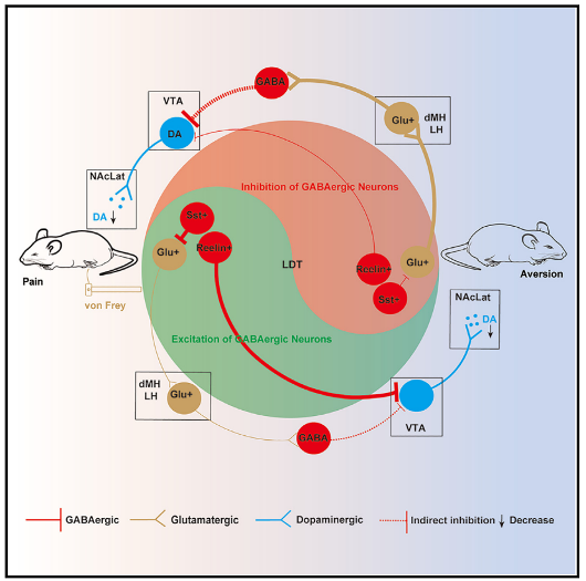
      Digitizing the study of neural circuits and naturalistic mouse beh...

    • Speaker:Kexin Yuan

      Institution:Tsinghua University

      Time:2023.10.12 10:00AM

      Locatiom:Meeting Room 205

      Unravelling the Connectivity, Function and Working Mechanisms of ...

    • Speaker:Rurong Ji

      Institution:Duke University

      Time:2023.10.11 9:00AM

      Locatiom:Meeting Room 205

      Regulation of pain, anesthesia, and cognition by immune checkpoint...

    • Speaker:Dr. Gilbert Kirouac

      Institution:University of Manitoba

      Time:2023.9.25 10:00AM

      Locatiom:705 Meeting Room

      Input-output organization of the paraventricular nucleus of the th...

    • Speaker:Liqun Luo

      Institution:Stanford University

      Time:2023.9.7 15:45

      Locatiom:Liangzhu Laboratory

      Wiring Specificity of Neural Circuits

      • FACULTY

        FACULTY

        VIEW MORE

        School of Brain Science and Brain Medicine Zhejiang University

        The School of Brain Science and Brain Medicine, devoted to the study of neuroscience and neuromedicine, was founded in October 2019. As the first school focusing on brain science and brain medicine in Chin... 【More】